Understanding Ultrasound: A Simple Guide with Procedure, When It’s Needed, and Daily Life Tips After Diagnosis
The medical procedure of ultrasound provides internal body imaging using high-frequency sound waves without causing pain to patients. This non-invasive medical test offers safe care for pregnant women because it operates without exposing patients to radiation. Doctors frequently prescribe ultrasound testing to identify internal anatomy and analyze bodily tissue as well as evaluate blood circulation patterns for diagnosing or clinical monitoring purposes. The following blog contains information about ultrasound technology including its operation and physician recommendations and required patient modifications after the examination.
What Is an Ultrasound and How Does It Work?
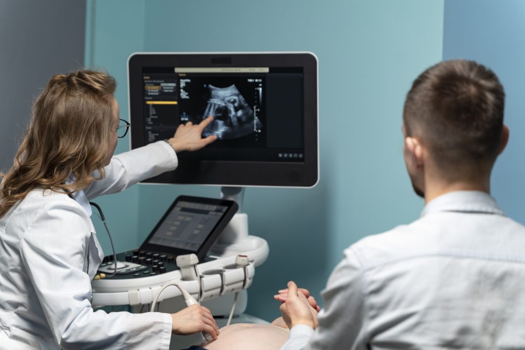
A sonogram or ultrasound produces detailed images of internal body structures by transmitting sound waves through the body. Universal ultrasound exams utilize sound waves instead of radiation to create images while remaining safer than X-ray tests. The technician applies skin-preparing gel before operating the transducer device to conduct the examination. During an ultrasound procedure the transducer directs sound waves through the body to create images which display on a screen through echo detection from the body tissue. Doctors can visualize internal body processes using an imaging technique that enables them to observe without surgery or causing harm. Ultrasound testing requires 15 to 30 minutes to complete while it produces no adverse effects on the body.
A specialty type of ultrasound known as Doppler can assess blood flow through vessels.
Why Do Doctors Recommend an Ultrasound?
- Doctors usually recommend an ultrasound when they need to:
- Investigate the cause of pain, swelling, or infection in the body
- Check the condition of organs like the liver, kidneys, gallbladder, spleen, and pancreas
- Monitor the growth and development of a baby during pregnancy
- Examine lumps or abnormalities in soft tissues
- Guide doctors during procedures like needle biopsies
- Diagnose heart conditions with echocardiography
- Assess blood flow in arteries and veins (Doppler ultrasound)
Ultrasound remains the preferred initial diagnostic procedure during situations when a patient or healthcare provider feels unexplained discomfort. The technology offers immediate secure data output which supports medical professionals to make instant clinical decisions.
Common Situations When an Ultrasound is Needed
Abdominal Pain: Doctors perform abdominal ultrasounds to detect persistent stomach pain which helps identify problems such as gallstones kidney stones liver disease and appendix inflammation.
Pregnancy: Monitoring baby development represents the main function of ultrasound tests because they also detect heartbeats and examine the placenta while determining fetal positioning.
Pelvic Issues: Pelvic ultrasonography examines female reproductive organs to detect abnormal periods or fibroids or cysts.
Heart Concerns: Doctors use echocardiograms to examine how the heart works as well as locate valve problems or evaluate heart muscle performance.
Swollen Glands or Lumps: A doctor uses neck or breast ultrasound scans to examine unexplained swollen areas or lumps.
Blood Clots or Poor Circulation: During Doppler ultrasound doctors evaluate blood vessels to identify potential clots and blockages primarily in the legs.
Language Tip: Use of “As” in Medical Context
Medical conversations make frequent use of the word “as” to explain why things happen or what causes them. It often means “because” or “since.”
For example:
“As you are having lower abdominal pain, we are doing an ultrasound.”
“As your heart rate is irregular, an echocardiogram is recommended.”
“As you are in your second trimester, it’s time for a detailed pregnancy scan.”
Patients can better understand medical instructions through knowledge of these simple words.
What to Expect During the Ultrasound Procedure
Ultrasound testing takes place inside hospital facilities alongside diagnostic centers and clinics. You will need to get dressed in a gown before assuming a comfortable position on the examination table. Before performing abdominal scans medical staff will request patients to fast for 8-12 hours. The technician will ask you to drink water before pelvic ultrasound because a full bladder enhances imaging quality.
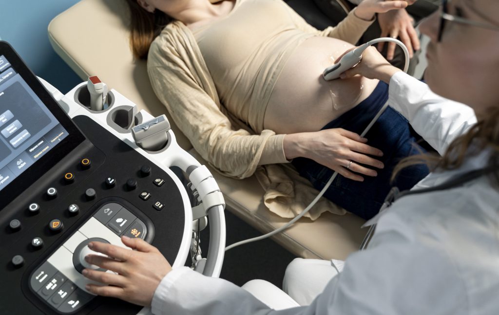
The sonographer will place gel on your skin to create a smooth interface before using the transducer. The diagnostic equipment operator starts moving the transducer over the area of interest. The pressure you feel during the examination will be very light and without pain. The diagnostic procedure executes without complications and takes no more than thirty minutes to finish.
The doctor permits most patients to leave the healthcare setting and continue with their regular routines until patients receive explicit instructions.
Daily Life Precautions After a Concerning Ultrasound
The particular medical condition discovered through ultrasound testing determines which specific precautions you must follow. However, here are general lifestyle tips you may be advised to follow:
Adjust Your Diet: You should avoid consuming processed and oily and salted foods. Your diet should contain more vegetables together with fruits and whole grains and lean protein sources. Drink plenty of fluids while possibly switching to green tea based on medical recommendations.
Follow-Up Appointments: Always honor your medical check-up appointments and follow-up test schedules. Such tests serve to track your development alongside possible modifications to your treatment plan.
Take Medications Properly: Always follow your doctor’s prescription and maintain the full recommended medication period regardless of symptom recovery.
Avoid Strain: Consult your doctor after getting an ultrasound that shows organ abnormalities such as enlarged liver or kidney stones because you must stay away from weightlifting and bodies-stressing movements.
Exercise Wisely: Your doctor will determine if you can perform light physical exercises based on your recovery status. A light walk can help your blood circulate while your body heals itself.
Stay Informed: Get your doctor to decode the report terminology and explain its findings. Learning about your medical situation provides peace of mind while increasing your ability to handle it well.
Summary Table: When and Why You Might Need an Ultrasound
| Reason | Ultrasound Purpose |
| Abdominal pain | Detect gallstones, liver issues, or appendix problems |
| Pregnancy check | Monitor baby’s growth and detect complications |
| Irregular periods or pelvic pain | Examine uterus, ovaries, and reproductive health |
| Unusual swelling or lumps | Identify cysts, tumors, or swollen glands |
| Heart irregularities | Check heart structure and pumping through echocardiogram |
| Blood circulation problems | Detect blood clots and assess blood flow (Doppler) |
Final Thoughts: Why You Shouldn’t Ignore Ultrasound Advice
Ultrasound serves multiple functions beyond its use for pregnancy monitoring. This diagnostic tool identifies health problems in early stages when symptoms are still manageable. Ultrasound proves essential for diagnostics by helping doctors examine organs and make sense of chronic pain while assisting in surgical guidance procedures.
Once your doctor suggests an ultrasound test you should not delay booking an appointment. The procedure stands as a secure method that poses no pain while immediately revealing internal body specifics. Taking necessary precautions regarding diet and activity while following up properly after a diagnosis will significantly improve your health outcomes.